In short: Full-body photo documentation is mainly worthwhile if you have a large number of moles or an increased risk of melanoma. Overview photos record where your moles are located. Digital dermoscopy can additionally track subtle changes in a selected mole over time. If a new or rapidly growing spot worries you, have it examined promptly, even if no comparison photos exist yet.
What do overview and close-up images show?
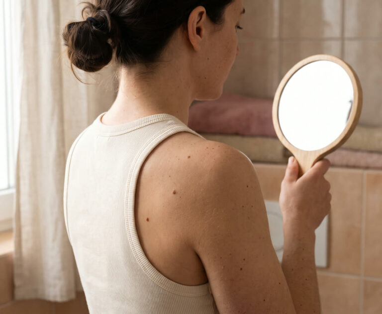
Imagine you notice a dark dot near your shoulder blade while getting changed. You can hardly see your own back. Was the dot there before? On an overview photo from your last skin check, the doctor can locate the spot using the neighbouring moles. If the skin there was clearly visible, she can check whether the dot was already present at the time. How it should be assessed medically today is decided by the examination.
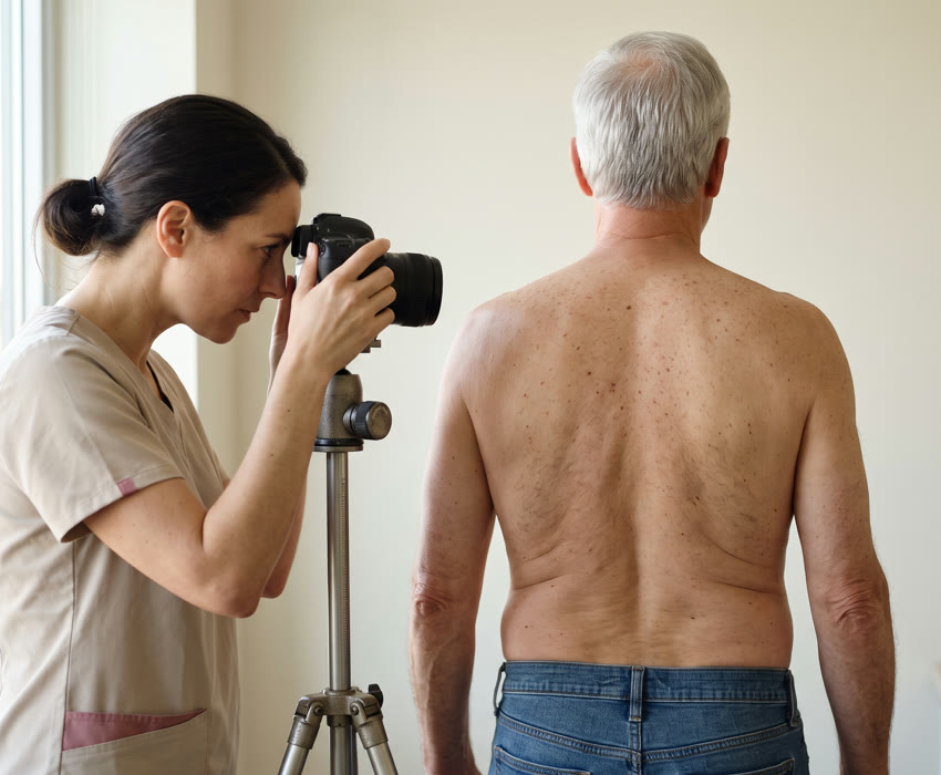
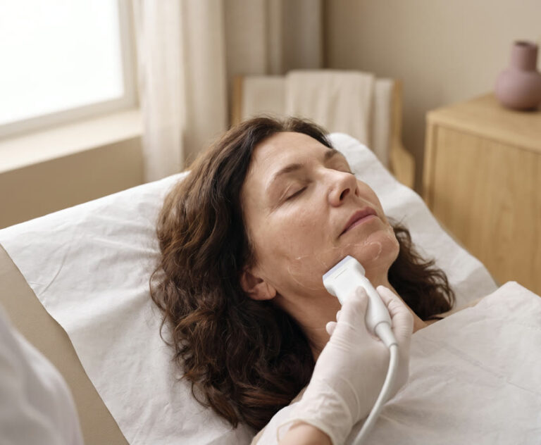
In total body photography, images of the visible skin are taken from defined angles. Together they form a photographic skin map, also known as «body mapping». It also covers areas of skin where no mole stood out at the first appointment. This matters because black skin cancer, melanoma, can also develop on skin that previously looked normal.
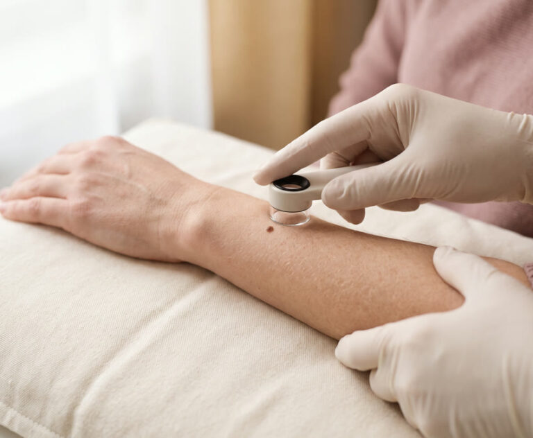
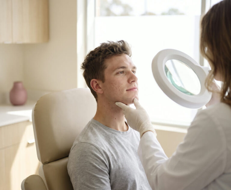
In dermoscopy, also called epiluminescence microscopy, the doctor examines a single mole with light and magnification. This allows her to see, for example, how the brown pigment is distributed within the mole. If she saves this view digitally, she can later record the same mole again under magnification. «Sequential» in this context means that images taken at different times are compared with each other.
Who benefits from photo documentation?
Whether images add value depends on your skin and your medical history. With a very large number of moles, it can be difficult to spot a new one among those already known, even during a careful examination. Several unusual-looking moles can also make a comparison useful. If you have had melanoma before, or if several close relatives have had it, mention this at your mole check in Zurich. How often the checks themselves make sense is explained in our article How often should you have your moles checked?
| Your situation | What helps you |
|---|---|
| You have many similar-looking moles, especially on your back. | Ask about overview photos. They make it easier to find a mole again and to detect new spots. |
| During the examination, a single mole stands out that the doctor considers suitable for monitoring. | A dermoscopic close-up records its current pattern. Agree on when exactly this spot will be assessed again. |
| You have few unremarkable moles and no known particular risk factors. | An ongoing photo series of the whole body is usually not necessary in this situation. You can show a specific change at any time. |
| A spot has changed noticeably within a short time. | Have it examined promptly. You need neither an older photo nor an appointment for a full-body series. |
You do not need to count your moles before the appointment. It is more helpful if you can say which spot caught your attention and since when. The doctor can then discuss with you whether an image of that spot is enough or whether photos of larger skin areas would be useful for later checks.
Photos do not replace an early appointment
Photo documentation is meant for monitoring over time. If a spot appears, changes in shape or colour, itches or bleeds, do not wait for the next scheduled imaging. Arrange an examination promptly.
How are the comparison images taken?
At the first examination, you discuss your medical history and show any spot that concerns you. For the overview photos, you uncover the areas of skin to be recorded. Jewellery or hair can hide a spot. Ask beforehand which areas will be in the picture. If you would prefer to keep a part of your body covered, you can discuss how the skin there can be examined separately.
Depending on the method, you stand in set positions for the images. Even the position of your arms affects how much skin on the sides of your body is visible. At a later session, the same areas are easier to compare if posture, angle and lighting are as similar as possible. The doctor can also take detailed images of selected moles and note which part of the body they belong to.
At the follow-up appointment, the doctor first finds the same spot on the earlier image. Has a new mole appeared? Has the distribution of colour within a known spot changed? She assesses such a difference together with today’s skin findings. A change can be benign. Depending on the findings, further monitoring is enough or the doctor recommends having tissue examined.
Where does the skin map have blind spots?
An overview only reaches as far as the camera can see. The hairy scalp, the soles of the feet and the skin under a breast or in a skin fold may be missing from the standard views. If you have noticed something exactly there, point to it. The doctor can then look at the spot directly and decide whether an additional image is useful.
Even a sharp close-up does not show individual tumour cells. If the examination raises suspicion of black skin cancer, the tissue must be examined in a laboratory. For this, the suspicious mole is removed completely where possible. This applies even if no difference can be seen on older photos. Conversely, a conspicuous photo can lead to a removal in which the finding turns out to be benign.
What does the clinic in Zurich offer?
At the Swiss Derma Clinic in Zurich, we examine the entire skin and assess moles with a dermatoscope. With digital video dermoscopy, we record selected pigmented moles and conspicuous skin changes. We store the images in your patient file and compare them with the current findings at follow-up examinations.
Our services for skin cancer screening in Zurich also include digital body mapping with total body photography. Software can point out new or changed spots. The medical assessment is carried out by our dermatologists. Which form of documentation makes sense for you is something we discuss on the basis of your personal risk profile.
Frequently asked questions
What is the first image for if there is no older one?
It records the baseline findings. The doctor can already assess a mole with the dermatoscope at this appointment. If she selects it for monitoring, it is later possible to look specifically at how its pattern looked at the first visit.
What should I ask about data protection before the images are taken?
If full-body photos feel too personal, raise this before the images are taken. You can ask which views are needed for your situation, where the photos are stored and who can access them for comparison.
How often are the images repeated?
That depends on your personal risk and the findings. Your doctor agrees the intervals with you. A single mole that is being monitored specifically may be recorded again after just a few months. Overview images of the whole body are usually repeated at longer intervals.
Can I bring my own photo to the appointment?
Yes. If you have photographed the spot before, show the picture and give the date it was taken if possible. It can help to identify the spot. However, different lighting and distances make it harder to compare colour and size. The doctor assesses the fine structures with the dermatoscope.
What should I do if I notice something before the check-up?
Get in touch promptly if you discover a new unusual spot or a known spot grows noticeably. Mention the reason when booking the appointment. This also applies if the spot was unremarkable at the last check-up or has not yet appeared on any image.
This article does not replace a medical examination.