In brief
The so-called white skin cancer often manifests itself as a non-healing skin change. The areas are often nodular, scaly or crusty. Sometimes they present as wounds that refuse to heal. At the Swiss Derma Clinic, the diagnosis is made by clinical examination and, if necessary, dermoscopy. Treatment depends on the type, size and location of the tumor and in many cases consists of complete surgical removal.
Basal cell carcinoma and squamous cell carcinoma
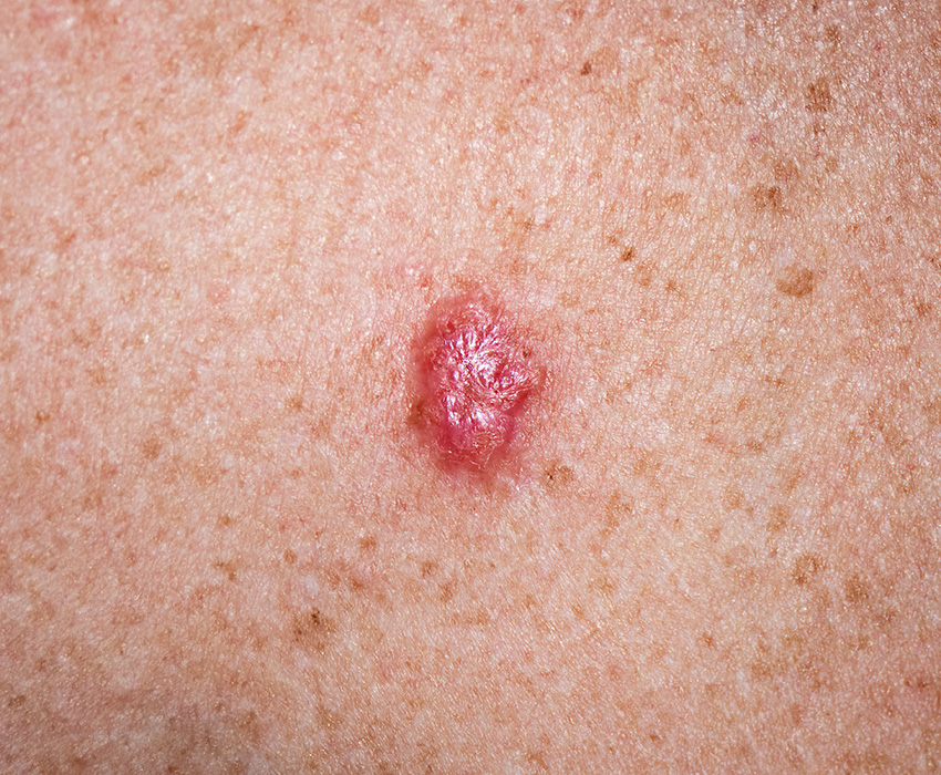
Basal cell carcinoma usually grows slowly and rarely forms metastases. It often presents as a skin-colored or slightly shiny lump, sometimes with small visible vessels. Flat, scar-like changes are also possible and should be clarified by a specialist.
Squamous cell carcinoma often develops from actinic keratoses, so-called skin cancer precursors. It is characterized by hardened, scaly or crusted areas of skin that visibly change or do not heal.
Both forms can usually be treated well. The prerequisite: they should be recognized as early as possible.
What are the typical signs of white skin cancer?
White skin cancer usually develops gradually over a period of many years. Certain skin types with very light skin are particularly at risk. Those affected often notice open, inflamed, crusty or altered areas of skin that persist for weeks or months.
Indications can be:
- Skin areas that do not heal
- Recurrent crusts or bleeding
- Rough, scaly or hardened areas
- Newly appearing lumps or thickenings
- Changes to existing skin areas
Such changes are often underestimated, especially on the face. Such skin changes can, but do not necessarily have to be indicative of white skin cancer - an early specialist examination ensures safety.
The dermatological examination for suspected white skin cancer
We always start with a clinical examination. The skin is systematically assessed by a specialist.
With the help of dermoscopy, we can also recognize structures that are not visible to the naked eye. This examination also provides us with important information about the type of change.
If the findings are unclear or a tumor is suspected, we will remove the skin change or take a tissue sample. The subsequent histological examination then confirms the diagnosis. We will then discuss the next steps together.
Removal of the skin change and further planning
The treatment depends on the type, size and location of the tumor. In most cases, the skin change is surgically removed - this is often possible on an outpatient basis in our practice in Zurich. The tumor is removed with a safety margin. The distance is important to prevent recurrence at this site.
Depending on the findings, additional procedures may be useful. For example, in the case of very superficial forms or certain localizations. The decision is always based on the individual findings and the current medical guidelines.
After removal, the tissue is examined histologically. This examination provides the basis for further planning.
Course and aftercare
After successful treatment, regular check-ups are necessary.
Follow-up care serves to detect new skin changes at an early stage. The check-up intervals depend on the individual risk.
Prevention: UV protection is important!
Long-term UV exposure is the most significant risk factor for white skin cancer. Sun protection plays a central role in prevention and aftercare.
The recommendation:
- Daily UV protection with a suitable sun protection factor
- Protect yourself with suitable clothing and headgear, especially if you have thin or thinning hair
- Avoid intensive sunbathing or visits to the solarium
Regular skin checks help to detect changes at an early stage. Make an appointment for this at the Swiss Derma Clinic in Zurich.
Your specialists in Zurich
At the Swiss Derma Clinic, experienced dermatology specialists are at your side. We can safely assess your skin changes and offer you a treatment that is individually tailored to you.
Do you have a skin lesion that is not healing or is causing you concern?
Then have your skin examined by a specialist - at the Swiss Derma Clinic in Zurich.